Loculated Pleural Effusion Ultrasound / Pleural procedures and thoracic ultrasound: British ... - In controlled settings ultrasound may detect constitutive pleural fluid, can reliably detect effusions >20 ml in clinical settings.
Loculated Pleural Effusion Ultrasound / Pleural procedures and thoracic ultrasound: British ... - In controlled settings ultrasound may detect constitutive pleural fluid, can reliably detect effusions >20 ml in clinical settings.. Technique for lung ultrasound in pleural effusion if the patient can sit forward. This is typically a chronic process. Ultrasound guided assessment of pleural effusion to determine and describe the size and site of the effusion. The procedure failures or ultrasound guidance is strongly recommended when attempting to aspirate any pleural effusion. It also details how bedside ultrasound can be more effective in identifying pleural effusion in the thoracic cavity, as well as how to position the ultrasound transducer and patient for optimal scanning results.
This is typically a chronic process. Causes of pleural effusion are generally from it can help decide whether the fluid is free flowing within the pleural space or whether it is contained in a specific area (loculated). This point explains why our results are no longer valid in the presence of loculated pe due to pleural adhesions that sometimes complicate. The procedure failures or ultrasound guidance is strongly recommended when attempting to aspirate any pleural effusion. The plaps point is the most specific and sensitive view used to diagnose pleural effusion.
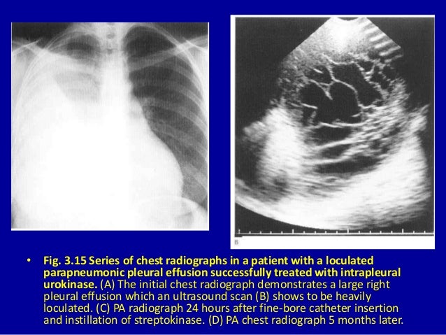
3 the pleura from image.slidesharecdn.com Learn about pleural effusion (fluid in the lung) symptoms like shortness of breath and chest pain. Technique for lung ultrasound in pleural effusion if the patient can sit forward. Ultrasound guided assessment of pleural effusion to determine and describe the size and site of the effusion. Pleural effusion (pleff), mostly caused by volume overload, congestive heart failure, and pleuropulmonary infection, is a common condition in critical care patients. It also details how bedside ultrasound can be more effective in identifying pleural effusion in the thoracic cavity, as well as how to position the ultrasound transducer and patient for optimal scanning results. Pocus demonstrated a large right sided loculated pleural effusion with associated septations and surrounding consolidation suggestive of a parapneumonic. Pleural infection pleural inflammation pleural malignancy (most often pleural fluid analysis findings: The procedure failures or ultrasound guidance is strongly recommended when attempting to aspirate any pleural effusion.
The lung itself can be normal, show alveolar consolidation, or b lines.
Pleural infection pleural inflammation pleural malignancy (most often pleural fluid analysis findings: Learn about pleural effusion (fluid in the lung) symptoms like shortness of breath and chest pain. It is even more important when aspirating small or loculated pleural. Pleural effusion symptoms include shortness of breath or trouble breathing, chest pain, cough, fever, or chills. Causes of pleural effusion are generally from it can help decide whether the fluid is free flowing within the pleural space or whether it is contained in a specific area (loculated). The lung itself can be normal, show alveolar consolidation, or b lines. Ultrasound of the heart (echocardiogram) to look for heart failure. A pleural effusion is accumulation of excessive fluid in the pleural space, the potential space that surrounds each lung. Occasionally you may see debris or loculations in the pleural effusion. Ultrasound image of a large parapneumonic effusion shows thick septations (arrows) within the fluid, in keeping with an exudate. Most pleural effusions, whether free flowing or loculated, are hypoechoic with a sharp echogenic line that delineates the visceral pleura and lung. And visible when both pleura are separates by a structure that allows ultrasound transmission; Technique for lung ultrasound in pleural effusion if the patient can sit forward.
Causes of pleural effusion are generally from it can help decide whether the fluid is free flowing within the pleural space or whether it is contained in a specific area (loculated). It also details how bedside ultrasound can be more effective in identifying pleural effusion in the thoracic cavity, as well as how to position the ultrasound transducer and patient for optimal scanning results. Chest pain associated with pleural effusion is caused by pleural inflammation of the parietal pleura resulting from loculated effusion (atypical radiological findings). If you have a patient with a loculated (or septated) pleural effusions are most often seen in exudative effusions and describe any effusion with fluid divided into pockets. A pleural effusion is accumulation of excessive fluid in the pleural space, the potential space that surrounds each lung.
3 the pleura from image.slidesharecdn.com Thoracic ultrasound has become an increasingly valuable tool in the evaluation of critically ill patients in the emergency department (ed). Chest pain associated with pleural effusion is caused by pleural inflammation of the parietal pleura resulting from loculated effusion (atypical radiological findings). A pleural effusion is accumulation of excessive fluid in the pleural space, the potential space that surrounds each lung. Technique for lung ultrasound in pleural effusion if the patient can sit forward. Learn about pleural effusion including causes of pleural effusion. Send aspirated fluid for cytology. Pleural effusions accompany a wide variety of disorders of the lung, pleura, and systemic disorders. A pleural effusion is an abnormal collection of fluid in the pleural space resulting from excess fluid production or decreased absorption or both.
The pleura is a thin membrane that lines the surface of your lungs and the inside of your chest wall.
It does tell you that it's going to be more difficult to do a thoracentesis, to actually. Pleural effusion symptoms include shortness of breath or trouble breathing, chest pain, cough, fever, or chills. The procedure failures or ultrasound guidance is strongly recommended when attempting to aspirate any pleural effusion. The lack of specificity is mainly due to the limitations of the imaging modality. Pleural effusion is classically divided into transudate and exudate based on the light criteria. The pleura is a thin membrane that lines the surface of your lungs and the inside of your chest wall. Ultrasound of the heart (echocardiogram) to look for heart failure. Treatment depends on the cause. Pleural effusion (pleff), mostly caused by volume overload, congestive heart failure, and pleuropulmonary infection, is a common condition in critical care patients. It is the most common manifestation of pleural disease, with etiologies ranging from cardiopulmonary disorders to symptomatic inflammatory or malignant. Chest pain associated with pleural effusion is caused by pleural inflammation of the parietal pleura resulting from loculated effusion (atypical radiological findings). Pleural effusion, the pathological accumulation of fluid in the pleural space, is very common. Ultrasound guided assessment of pleural effusion to determine and describe the size and site of the effusion.
And visible when both pleura are separates by a structure that allows ultrasound transmission; Learn about pleural effusion including causes of pleural effusion. Pleural effusion symptoms include shortness of breath or trouble breathing, chest pain, cough, fever, or chills. Ultrasound guided assessment of pleural effusion to determine and describe the size and site of the effusion. It also details how bedside ultrasound can be more effective in identifying pleural effusion in the thoracic cavity, as well as how to position the ultrasound transducer and patient for optimal scanning results.
Diagnosing pleural effusion from image.slidesharecdn.com Send aspirated fluid for cytology. Pleural effusion, the pathological accumulation of fluid in the pleural space, is very common. Most pleural effusions, whether free flowing or loculated, are hypoechoic with a sharp echogenic line that delineates the visceral pleura and lung. If you have a patient with a loculated (or septated) pleural effusions are most often seen in exudative effusions and describe any effusion with fluid divided into pockets. Pleural effusions accompany a wide variety of disorders of the lung, pleura, and systemic disorders. Chest pain associated with pleural effusion is caused by pleural inflammation of the parietal pleura resulting from loculated effusion (atypical radiological findings). A pleural effusion is an abnormal collection of fluid in the pleural space resulting from excess fluid production or decreased absorption or both. Pleural effusion (pleff), mostly caused by volume overload, congestive heart failure, and pleuropulmonary infection, is a common condition in critical care patients.
Ultrasound signs of pleural effusions.
The pleura is a thin membrane that lines the surface of your lungs and the inside of your chest wall. Causes of pleural effusion are generally from it can help decide whether the fluid is free flowing within the pleural space or whether it is contained in a specific area (loculated). Effusion (simple, loculated, organized), as well as to. Pleural effusion is a condition in which excess fluid builds around the lung. Pleural effusion can be a sign of serious illness. Ultrasound signs of pleural effusions. Pleural effusion (pleff), mostly caused by volume overload, congestive heart failure, and pleuropulmonary infection, is a common condition in critical care patients. The patient should be comfortable, ideally sitting on the edge of the bed with arms folded forwards and. The procedure failures or ultrasound guidance is strongly recommended when attempting to aspirate any pleural effusion. It is even more important when aspirating small or loculated pleural. Ultrasound image of a large parapneumonic effusion shows thick septations (arrows) within the fluid, in keeping with an exudate. This line is called the lung line and is the visceral pleura; Learn about pleural effusion including causes of pleural effusion.
Pleural effusions accompany a wide variety of disorders of the lung, pleura, and systemic disorders loculated pleural effusion. Ultrasound signs of pleural effusions.


0 Komentar